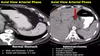
CT Scan Comparison: Normal Stomach vs Gastric Adenocarcinoma - Early and Advanced Stage Cancer Cases
Explore CT scan images comparing normal stomach to gastric adenocarcinoma, examining key features and differences in early and advanced cancer stages.
Correlation Lecture: Imaging vs Catheterization - Cardiovascular Diagnostics
Explore the correlation between imaging and catheterization in cardiovascular diagnostics. Gain insights into advanced techniques for accurate assessment and treatment planning in cardiology.
Role of CT in Evaluation of Right Ventricle
Explore CT's role in evaluating the right ventricle with Dr. Waleed Ahmed. Learn key techniques and insights for improved cardiovascular assessment and patient care.
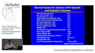
Evaluation of RV Size and Function by TTE
Explore advanced techniques for evaluating right ventricular size and function using transthoracic echocardiography in this comprehensive cardiovascular education session.
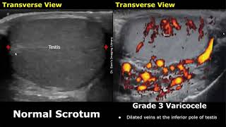
Varicocele Grading on Color Doppler Ultrasound - Grades 1-5
Learn to grade varicoceles using color Doppler ultrasound, from Grade 1 to 5. Explore key characteristics and diagnostic criteria for each grade through detailed case studies.

Uterus MRI - Normal vs Abnormal Images, Uterine and Cervix Pathologies, Cancer Staging in Gynecology
Explore uterine and cervical pathologies through MRI images, covering normal anatomy, various abnormalities, and cancer staging in gynecology.
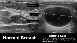
Small Parts Ultrasound - Normal vs. Abnormal Images for Thyroid, Breast, Scrotum, Testis, and Eye
Explore normal vs abnormal ultrasound images of small body parts including thyroid, breast, scrotum, eye, salivary glands, and adrenal glands to enhance diagnostic skills.
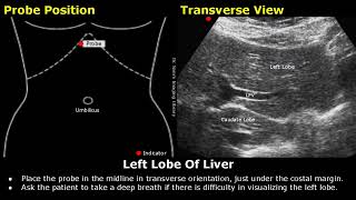
Abdominal, Gynecological and Obstetric Ultrasound Probe Positioning - Transducer Placement for USG Scan
Master ultrasound probe positioning for abdominal, gynecological, and obstetric scans. Learn precise transducer placement techniques for various organs and structures to enhance diagnostic accuracy.
Multi-Modality Case Presentations by Advanced CV Imaging Fellows - Lecture
Explore advanced cardiovascular imaging through multi-modality case presentations by CV imaging fellows. Gain insights into complex cardiac conditions and diagnostic techniques.
Imaging in Preventive Cardiology
Explore advanced imaging techniques in preventive cardiology with Dr. Khurram Nasir. Gain insights into multimodality approaches for early detection and risk assessment of cardiovascular diseases.
Multi-Modality Case Presentations by Advanced CV Imaging Fellows - Lecture
Explore advanced cardiovascular imaging through multi-modality case presentations by CV imaging fellows, guided by Dr. Stephen Little. Gain insights into complex cardiac conditions and diagnostic techniques.
Multi-Modality Imaging Artifacts - How to Recognize and Overcome
Explore multi-modality imaging artifacts in cardiovascular diagnostics, learning to recognize and overcome challenges for improved patient care and diagnostic accuracy.
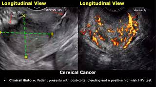
Cervix Ultrasound Reporting - Nabothian Cysts, Polyp, Fibroid, Cervical Incompetence, Cancer USG
Explore cervical ultrasound reporting, covering normal anatomy and various conditions including cysts, polyps, fibroids, incompetence, and cancer.
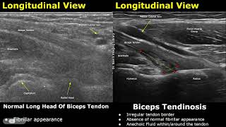
Elbow Ultrasound: Normal vs Abnormal Images - Muscles, Tendons, Ligaments and Nerves MSK USG
Explore normal vs. abnormal elbow ultrasound images, covering muscles, tendons, ligaments, and nerves. Learn to identify various conditions and injuries through detailed MSK USG examples.
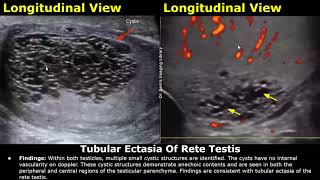
Scrotal Ultrasound Reporting - Scrotum, Testis and Epididymis Pathologies
Master scrotal ultrasound reporting techniques and learn to identify various pathologies of the scrotum, testis, and epididymis. Enhance your skills in writing comprehensive USG reports for accurate diagnoses.
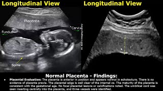
Placenta Ultrasound Reporting - How to Write Pregnancy USG Reports for Pathologies and Positions
Learn to interpret and report placental ultrasounds, covering normal anatomy, pathologies, and positioning. Master the skills to accurately assess and document various placental conditions during pregnancy.
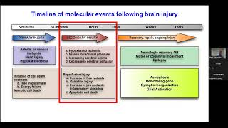
Pediatric Disorder Consciousness - Neuroprotection and Neuroprognostication
Explore neuroprotection and neuroprognostication in pediatric disorder consciousness with expert insights from Dr. Mark Wainwright.
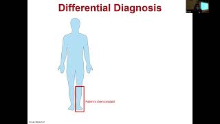
Case Records of the MGH - An 80-Year-Old Man with Foot Drop
Explore a medical case study of an elderly patient with foot drop, analyzing symptoms, diagnosis, and treatment options for this neurological condition.
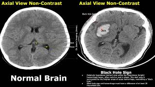
CT Scan Brain: Normal vs. Hemorrhagic Stroke - Identifying Key Signs
Explore CT scan images of normal brain vs hemorrhagic stroke. Learn to identify key signs like swirl, black hole, blend, spot, and island for accurate diagnosis and patient care.
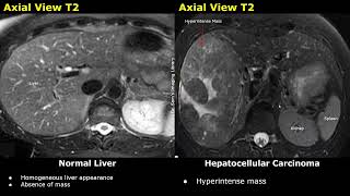
Liver MRI - Normal vs Hepatocellular Carcinoma (HCC) Images: T1, T2, DWI, ADC and Contrast Sequences
Explore MRI imaging of normal liver vs hepatocellular carcinoma. Learn to identify HCC on T1, T2, DWI, ADC, and contrast sequences. Understand causes, risk factors, and diagnostic markers.
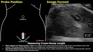
Step-by-Step Guide on 7 Week Pregnancy Transabdominal Ultrasound Scan - Full Protocol with Reporting
Learn to perform and interpret a 7-week pregnancy transabdominal ultrasound scan, covering patient positioning, measurements, fetal heart rate assessment, and reporting protocols.