Multi-Modality Case Presentations by Advanced CV Imaging Fellows - Lecture
Explore advanced cardiovascular imaging through multi-modality case presentations by CV imaging fellows, guided by Dr. Stephen Little. Gain insights into complex cardiac conditions and diagnostic techniques.
Multi-Modality Imaging Artifacts - How to Recognize and Overcome
Explore multi-modality imaging artifacts in cardiovascular diagnostics, learning to recognize and overcome challenges for improved patient care and diagnostic accuracy.
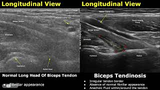
Elbow Ultrasound: Normal vs Abnormal Images - Muscles, Tendons, Ligaments and Nerves MSK USG
Explore normal vs. abnormal elbow ultrasound images, covering muscles, tendons, ligaments, and nerves. Learn to identify various conditions and injuries through detailed MSK USG examples.
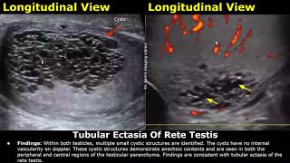
Scrotal Ultrasound Reporting - Scrotum, Testis and Epididymis Pathologies
Master scrotal ultrasound reporting techniques and learn to identify various pathologies of the scrotum, testis, and epididymis. Enhance your skills in writing comprehensive USG reports for accurate diagnoses.
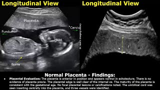
Placenta Ultrasound Reporting - How to Write Pregnancy USG Reports for Pathologies and Positions
Learn to interpret and report placental ultrasounds, covering normal anatomy, pathologies, and positioning. Master the skills to accurately assess and document various placental conditions during pregnancy.
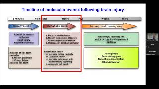
Pediatric Disorder Consciousness - Neuroprotection and Neuroprognostication
Explore neuroprotection and neuroprognostication in pediatric disorder consciousness with expert insights from Dr. Mark Wainwright.
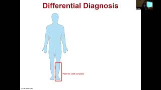
Case Records of the MGH - An 80-Year-Old Man with Foot Drop
Explore a medical case study of an elderly patient with foot drop, analyzing symptoms, diagnosis, and treatment options for this neurological condition.
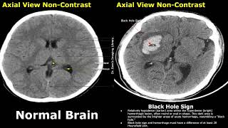
CT Scan Brain: Normal vs. Hemorrhagic Stroke - Identifying Key Signs
Explore CT scan images of normal brain vs hemorrhagic stroke. Learn to identify key signs like swirl, black hole, blend, spot, and island for accurate diagnosis and patient care.
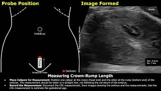
Step-by-Step Guide on 7 Week Pregnancy Transabdominal Ultrasound Scan - Full Protocol with Reporting
Learn to perform and interpret a 7-week pregnancy transabdominal ultrasound scan, covering patient positioning, measurements, fetal heart rate assessment, and reporting protocols.
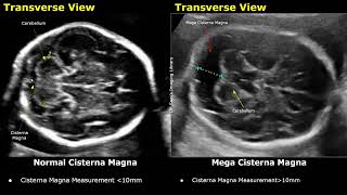
Edwards Syndrome (Trisomy 18) Antenatal Ultrasound Features - Normal vs Abnormal Fetal Images
Explore antenatal ultrasound features of Edwards Syndrome, comparing normal and abnormal fetal images across 16 key conditions for comprehensive understanding of Trisomy 18 diagnosis.
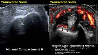
Wrist, Hand and Finger Ultrasound - Normal vs. Abnormal Images in Tenosynovitis and Rheumatoid Arthritis
Explore normal vs abnormal ultrasound images of wrist, hand, and finger conditions, including tenosynovitis and rheumatoid arthritis. Compare healthy structures with pathological findings for improved diagnostic skills.
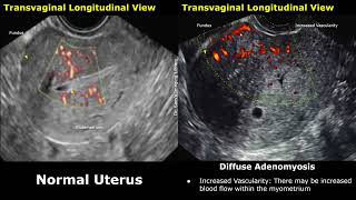
Uterus Ultrasound: Normal vs Adenomyosis Images - Diffuse and Focal Types - Gynecological USG Scan
Explore ultrasound imaging of normal uterus vs. adenomyosis, including diffuse and focal types. Learn key features for accurate gynecological diagnosis and differentiation.
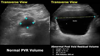
Pre Void and Post Void Residual Volume Bladder Ultrasound - Normal vs Abnormal Images
Learn to measure pre-void and post-void residual bladder volumes using ultrasound. Understand normal and abnormal results, calculation methods, and their clinical significance.
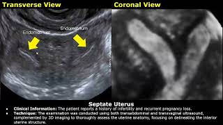
Congenital Uterine Anomalies Ultrasound Reporting - Didelphys, Bicornuate, Unicornuate Uterus
Explore congenital uterine anomalies through ultrasound reporting. Learn to identify and describe didelphys, bicornuate, unicornuate, septate, and arcuate uterus variations using USG techniques.
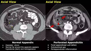
Appendix CT Scan: Normal vs Appendicitis - Acute, Gangrenous, and Perforated Types
Explore CT scan images of normal appendix vs. appendicitis, including acute, gangrenous, and perforated types. Learn key signs and characteristics for accurate diagnosis.
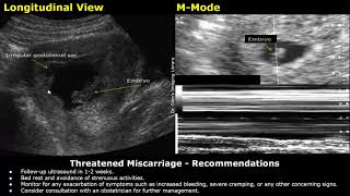
Miscarriage Ultrasound Reporting - Threatened, Missed, Inevitable, Incomplete Types USG Scan Reports
Learn to interpret and report ultrasound findings for various types of miscarriages, including threatened, missed, inevitable, incomplete, and complete.
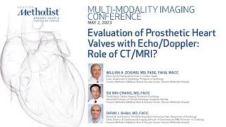
Multimodality Conference: Advances in Cardiovascular Imaging - 5.2.2023
Explore advanced cardiovascular imaging techniques with leading experts. Gain insights into multimodality approaches for improved diagnosis and patient care in cardiology.
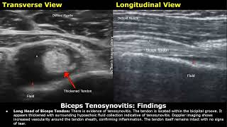
Shoulder Ultrasound Reporting - Supraspinatus Tendinosis, Tears, Biceps Tendon, and Joint Effusions
Master shoulder ultrasound reporting techniques for various conditions, including supraspinatus tendinosis, tears, biceps tendon issues, and joint effusions. Enhance diagnostic skills through case-based learning.
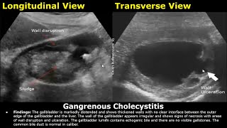
Abdominal Ultrasound Reporting - Liver, Gallbladder, Bile Duct, Pancreas, Spleen, Kidney, and Other Organs
Master abdominal ultrasound reporting techniques for liver, gallbladder, bile duct, pancreas, spleen, kidney, small bowel, hernia, appendix, prostate, and FAST examinations.
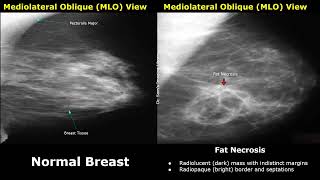
Mammography - Normal vs Abnormal Images and BI-RADS Classification for Breast Cancer and Other Diseases
Explore mammography images, distinguishing normal from abnormal findings. Learn BI-RADS classification and identify various breast conditions, from benign changes to malignancies.
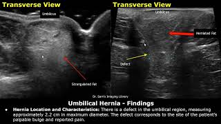
Hernia Ultrasound Reporting - Direct, Indirect, Inguinal, Epigastric, and Femoral Hernia USG Reports
Master ultrasound reporting for various hernia types, including epigastric, inguinal, femoral, and more. Learn to identify and describe key features for accurate diagnosis and effective communication.