Description:
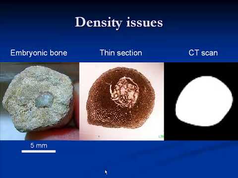
Explore the fascinating intersection of paleontology and modern imaging technology in this 59-minute Royal Tyrrell Museum Speaker Series talk. Delve into the history and applications of X-ray imaging in vertebrate paleontology, presented by Dr. Francois Therrien. Learn about various X-ray techniques, their challenges in fossil analysis, and how they overcome issues like fossilization changes and density variations. Discover the diverse uses of X-ray imaging, from assessing fossil presence to studying internal structures of ancient specimens. Examine intriguing case studies, including the analysis of dinosaur "hearts," fossilized gravid turtles, and elephant bird eggs. Gain insights into functional studies of dinosaur airways, brains, and inner ears, and understand how 3D rendering and finite element analysis contribute to paleontological research. Uncover the educational potential of X-ray imaging in bringing prehistoric life to light.
When X-Rays and Dinosaurs Collide: X-Ray Imaging in Vertebrate Palaeontology
Add to list