Description:
Explore a comprehensive 18-minute video comparing normal and abnormal neonatal brain ultrasound images for full-term infants and premature newborns. Delve into various conditions including hydrocephalus, germinal matrix hemorrhage, frontal horn cyst, and Chiari II malformation. Examine ultrasound findings for agenesis of corpus callosum, choroid plexus cysts, and lipomas. Learn to identify Dandy Walker malformation, mega cisterna magna, and septo-optic dysplasia. Understand the imaging characteristics of holoprosencephaly, schizencephaly, lissencephaly, and porencephaly. Recognize hydranencephaly, cystic encephalomalacia, and aqueductal stenosis on ultrasound. Study acute cerebellar hemorrhage, subarachnoid hemorrhage, and periventricular leukomalacia. Conclude with cerebral edema, ventriculitis, and meningitis, enhancing your ability to differentiate normal from pathological findings in neonatal brain ultrasounds.
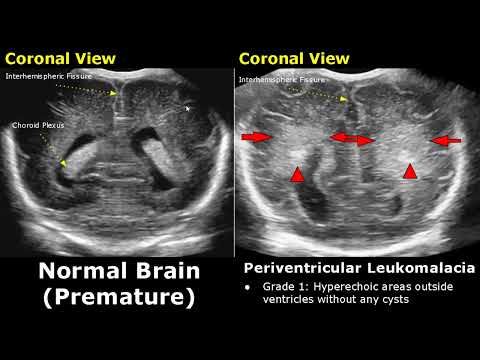
Neonatal Brain Ultrasound - Normal vs Abnormal Images in Full Term and Premature Infants
Add to list